Science is stunning


This year’s finalists for the Wellcome Image Awards
By Haley Hamblin From Mashable
It’s the 20th anniversary of the Wellcome Images Awards, which celebrates images of science and medicine. Their winners’ gallery features the most diverse range of images in the contest’s history. Anyone can submit to the collection, which began in 1997. The variety of images and methods of capturing them has expanded greatly over the last two decades. The awards celebrate the scientists, clinicians, artists and photographers who have contributed to bringing science to life through photos, illustrations and other imagery.
The overall winner and additional awards will be announced on March 15, 2017. Starting March 16, 2017, all images will be on display in the UK and around the world.
Unravelled DNA in a human lung cell
In order for plants and animals to grow and remain healthy, cells need to have the ability to replicate. During cell division, also known as mitosis, the entire DNA content of the cell is copied, with half going to each new cell. DNA is found in a region of the cell called the nucleus, which acts a bit like the brain. This picture shows the nucleus of one of two new daughter cells. The DNA in this cell has somehow become caught, and is being pulled between the two cells. This has caused the DNA to unfold inside the nucleus, and DNA fibres can be seen running through it. As the new cells have moved apart, the tension distributed by the rope-like DNA has deformed the nucleus’s usually circular envelope.
EZEQUIEL MIRON, UNIVERSITY OF OXFORD
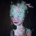
‘Hidden Learning’, from the Chrysalis project
Hidden Learning is taken from Chrysalis, a project at the University of St. Andrews designed to bring together women scientists at all stages of their careers to talk about issues and to seek advice and inspiration. Some of its conversations were interpreted by artist Sophie McKay Knight to create a body of work that was displayed in the Byre Gallery in St. Andrews as part of the Women in Science Festival 2016. A key aim of the Chrysalis project is to examine how creativity and imagination are required as well as integrity and precision when pursuing scientific research. Hidden Learning explores what women feel they keep hidden in the work environment, such as the pull between their career and home/other life – unique as that is to every woman. The veil seen in this image is made up of the molecular structure of a sugar molecule, contributed by one of the participating scientists.
ORIGINAL PAINTING BY SOPHIE MCKAY KNIGHT, WITH IMAGERY CONTRIBUTED BY WOMEN SCIENTISTS FROM THE UNIVERSITY OF ST ANDREWS – PART OF THE CHRYSALIS PROJECT COORDINATED BY MHAIRI STEWART
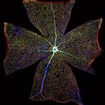
Surface of a Mouse Retina
The retina, located at the back of the eye, contains light-sensitive cells responsible for converting light into electrical nerve signals that the brain can process. As a result of aging or injury the retina can lose this function, causing vision loss. This image was created by digitally stitching together over 400 images to form one large image, so as to show the entire surface of a mouse retina. Blood vessels (blue) can be seen radiating from the centre of the image, supplying the entire retinal surface. Astrocytes, specialist cells of the nervous system, are double stained in red and green. These cells perform many functions – including maintaining and delivering nutrients to the nerves and the brain, and supporting the repair processes of the brain and spinal cord following injury – and are important for nerve survival and regeneration. Here, scientists are researching whether the function of astrocytes changes during retinal degeneration, which may lead to the development of new treatments for vision loss.
GABRIEL LUNA, NEUROSCIENCE RESEARCH INSTITUTE, UNIVERSITY OF CALIFORNIA, SANTA BARBARA
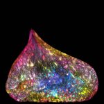
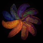
The Placenta Rainbow
The Placenta Rainbow highlights differences in mouse placental development that can result from manipulation of the mother’s immune system. These placentas were investigated at day 12 of the 20-day gestation period – the point at which a mouse’s placenta has gained its characteristic shape but is still developing. These placentas are from mice with genetically different immune systems, and have been stained for three proteins. Blue represents the nucleus, where DNA is stored and controlled; blood vessels are stained in red; and trophoblasts, the first cells to form in the developing embryo, are stained in green. Additional colors are present due to an expression of two or more of these proteins in the same cell. The range of colors indicates the significant effects that differences in a mother’s immune system can have on placental development. Such techniques could help us understand and identify ways to treat complications that arise during human pregnancies.
SUCHITA NADKARNI, WILLIAM HARVEY RESEARCH INSTITUTE, QUEEN MARY UNIVERSITY OF LONDON
Caricatural medieval medical practitioners
These scenes are inspired by works from medieval artists and the 15th-century Dutch painter Hieronymus Bosch. The scenes are separated by Asclepian snakes, representing both Asclepius – the ancient Greek god of medicine – and the modern-day symbol for medicine. Descriptions of the images (clockwise, from top left). 1) A parody of alchemy: a masked figure holds in his hand a conical flask from which frogs jump. In the distance a heron waits for a chance to grab one. 2) A man sits on a cart, asking a doctor if his disordered limbs can be repaired. 3) In an operation scene, a doctor appears to be pulling a bunch of sausages out of a patient’s belly while surrounded by hungry dogs. 4) A medieval surgeon holding a small knife operates on a man’s opened head. The patient is also being attended to by a female assistant, who is giving him some tea.
MADELEINE KUIJPER, MADELEINE KUIJPER ILLUSTRATIES
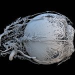
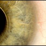
Vessels of a healthy mini-pig eye
A 3D model of a healthy mini-pig eye. The dent on the right-hand side of the image is the pupil, the opening that allows light into the eye. The blood vessels shown are bringing energy and food to the muscles surrounding the iris, which controls the amount of light entering the eye. The smallest vessels seen here are 20–30 micrometers (0.02–0.03 mm) in diameter. The other large vessels are feeder vessels for the retina, the light-sensing region at the back of the eye.
PETER M. MALOCA, OCTLAB AT THE UNIVERSITY OF BASEL AND MOORFIELDS EYE HOSPITAL, LONDON; CHRISTIAN SCHWALLER; RUSLAN HLUSHCHUK, UNIVERSITY OF BERN; SÉBASTIEN BARRÉ
Pigeon thermoregulation
All animals possess unique variations in their anatomy that help them adapt to their environment. Scott Echols is a member of the Grey Parrot Anatomy Project, which has been established to create technology that allows the world to study the anatomy of any animal. Discoveries made through the project have already been used when working with a large variety of animals, including humans. BriteVu, a novel contrast agent developed during the project, allows researchers to see the entire network of blood vessels in an animal, down to the capillary level. Images are taken from computed tomography scans. The intricate network of blood vessels in this pigeon’s neck is just visible at the bottom of the picture. This extensive blood supply just below the skin helps the pigeon control its body temperature through a process known as thermoregulation.
SCOTT ECHOLS, SCARLET IMAGING AND THE GREY PARROT ANATOMY PROJECT
Rita Levi-Montalcini
Rita Levi-Montalcini (1909–2012) was an Italian neurobiologist and the joint recipient of the 1986 Nobel Prize in Physiology or Medicine for the discovery of nerve growth factor (NGF). Rita graduated in medicine in 1936, but due to Mussolini’s 1938 Manifesto of Race, which barred non-Aryan citizens from having academic careers, was forced to build a small laboratory in the family home and work in secret. After the end of World War II she was invited to Washington University in St. Louis, USA, by Professor Viktor Hamburger, whose work was her inspiration. It was there that Rita discovered the role of NGF, which has increased the understanding of many conditions, including tumors, developmental malformations and dementia. Rita was the recipient of many awards, became a Foreign Member of the Royal Society and a United Nations Goodwill Ambassador, and was made an Italian senator for life. She also established the Rita Levi-Montalcini Foundation to support the education of girls and women in Africa.
DARIA KIRPACH/SALZMAN INTERNATIONAL
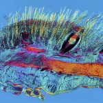
Cat Skin and Blood Supply
A polarised light micrograph of a section of cat skin, showing hairs, whiskers and their blood supply. This sample is from a Victorian microscope slide. Blood vessels were injected with a red dye called carmine dye (here appearing black) in order to visualize the capillaries in the tissue, a newly developed technique at the time. This image is a composite made up of 44 individual images stitched together to produce a final image 12 mm in width. Here, fine hairs (yellow), thicker whisker (yellow) and blood vessels (black) are all visible. Whiskers, unlike normal hair, are touch receptors, each containing a sensory organ called a proprioceptor. When a cat’s whiskers touch something, or feel vibrations in the air from a moving object, signals are sent from them to the brain to provide spatial awareness. Whiskers are therefore both a valuable hunting and survival tool.
DAVID LINSTEAD
Gabriel Galea, University College London
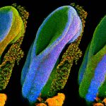
Our spines allow us to stand and move, and they protect the spinal cord, which connects all the nerves in our body with our brain. The spinal cord is formed from a structure called the neural tube, which develops during the first month of pregnancy. This series of three images shows the open end of a mouse’s neural tube, with each image highlighting (in blue) one of the three main embryonic tissue types. On the left is the neural tube itself, which develops into the brain, spine and nerves. On the right is the surface ectoderm – the word ‘ectoderm’ comes from the Greek ektos meaning ‘outside’ and derma meaning skin – which will eventually form the skin, teeth and hair. The middle image shows the mesoderm (also from Greek, meaning ‘middle skin’), which will form the organs. Problems can occur with neural tube development, such as in spina bifida, in which the bones of the spine and the spinal cord do not form correctly. Researchers are studying mouse neural tubes to try and prevent the development of such conditions.
GABRIEL GALEA, UNIVERSITY COLLEGE LONDON
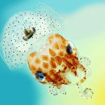
Hawaiian bobtail squid
Native to the Pacific Ocean, Hawaiian bobtail squid are nocturnal predators that remain buried under the sand during the day and come out to hunt for shrimp near coral reefs at night. The squid have a light organ on their underside that houses a colony of glowing bacteria called Vibrio fischeri. The squid provide food and shelter for these bacteria in return for their bioluminescence. The light organ is attached to an ink sac, which the squid uses like a type of shutter, controlling the amount of light released. The squid matches the light the bacteria produce to the moonlight and starlight, masking its silhouette and making it invisible to predators swimming below. This type of camouflage is called counter-illumination. This image shows a baby Hawaiian bobtail squid, measuring just 1.5 cm across. The black ink sac and light organ in the centre of the squid’s mantle cavity are clearly seen.
MARK R SMITH, MACROSCOPIC SOLUTIONS
Intraocular lens ‘iris clip’
This image shows how an ‘iris clip’, also known as an artificial intraocular lens (IOL), is fitted onto the eye. An iris clip is a small, thin lens made from silicone or acrylic material, and has plastic side supports, called haptics, to hold it in place. An iris clip is fixed to the iris through a 3 mm surgical incision, and is used to treat conditions such as myopia (nearsightedness) and cataracts (cloudiness of the lens). This particular patient, a 70-year-old man, regained almost full vision following his surgery.
MARK BARTLEY, CAMBRIDGE UNIVERSITY HOSPITALS NHS FOUNDATION TRUST
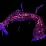
Language pathways of the brain
The brain is composed of two types of matter. Grey matter contains cells, and is responsible for processing information. White matter connects these areas of grey matter, allowing information to be transferred between distant areas of the brain. Areas responsible for speech and language have been mapped to two different brain regions. This image shows a 3D-printed reconstruction of the white matter pathway connecting these two areas (here shown from the left) which is called the arcuate fasciculus.
STEPHANIE J FORKEL AND AHMAD BEYH, NATBRAINLAB, KING’S COLLEGE LONDON; ALFONSO DE LARA RUBIO, KING’S COLLEGE LONDON
MicroRNA scaffold cancer therapy
Short genetic sequences called microRNAs, which control the proper function and growth of cells, are being investigated by researchers as a possible cancer therapy. However, their potential use is limited by the lack of an efficient system to deliver these microRNAs specifically to cancerous cells. Researchers at MIT have developed such a system, combining two microRNAs with a synthetic polymer to form a stable woven structure a bit like a net. This synthetic net can coat a tumor and deliver the two microRNAs locally to cancer cells. The two microRNAs used have different mechanisms of action and work together as a two-pronged attack: one is a tumor suppressor, and the other is an anti-microRNA, meaning that it prevents a mutated, tumor-promoting microRNA from functioning. This therapy has already been tested in mouse models of breast cancer, where it caused a tumor to shrink by nearly 90 percent after just two weeks.
JOÃO CONDE, NURIA OLIVA AND NATALIE ARTZI, MASSACHUSETTS INSTITUTE OF TECHNOLOGY (MIT)
Patient receiving treatment during outreach eye screening in India
A patient being treated by an eye doctor at a makeshift eye clinic in India. This image was taken while Susan, the photographer, was volunteering for the charity Unite For Sight. The charity has a long-term aim to improve global eye health and was founded in 2000. It has been responsible for providing over 90,000 cataract surgeries and giving eye care to 1.9 million people, some of whom live in extreme poverty. Clinics are set up in various locations, including schools. At this clinic, those requiring further treatment were taken to Kalinga Eye Hospital in Dhenkanal, Odisha, where they received eye surgery. They returned to their villages the following day.
SUSAN SMART
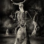
Stickman – The Vicissitudes of Crohn’s (Resolution)
This image is part of a series called Stickman – The Vicissitudes of Crohn’s. Its images are based around the character Stickman, a proxy or alter ego of the artist, who suffers from Crohn’s disease. He is made of sticks rather than bones and references the associated symptoms of weight loss, the body’s fragility following a flare-up, and the abrupt, transformative nature of Crohn’s. Here, Stickman is chastising his creator, the artist, for deriving artistic inspiration from his illness. However, this is ultimately an image of hope and regeneration, as suggested symbolically by the stick thrust into the earth and the hare in the tree’s womb. Crohn’s disease is a chronic condition caused by inflammation of the digestive system. Its cause has not yet been discovered, but scientists believe it is a combination of genetics, an abnormal reaction of the immune system to gut bacteria, and unknown environmental triggers, such as viruses, bacteria, stress or smoking.
SPOOKY POOKA
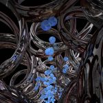
Synthetic DNA channel transporting cargo across membranes
Every cell is surrounded by a membrane, which serves to protect the cell’s contents from its external environment, provide support and connect the cell to others to form tissues and organs. Tube-like channels made of proteins span this membrane and control two-way communication between the cell and its environment. Researchers are using DNA as a building material to make synthetic channels that behave in exactly the same way. This image is an artist’s representation of what these channels look like. Cargo traveling through the channel is shown as colored spheres, while the mirrored coils around the edge of the image represent six DNA double helices, making up the walls of the channel. These DNA nanostructures are currently being engineered for use in vaccines, biofuels and biosensors and as research tools.
MICHAEL NORTHROP
Two young boys in rural Nicaragua
In Chichigalpa, a town in Nicaragua in Central America, chronic kidney disease (CKD) affects more than half of the adult population. One in three men are in the end stage of kidney (renal) failure, and CKD is responsible for 75 percent of deaths of men aged 35 to 55. At least 20,000 people are estimated to have died from the disease in the past two decades. Chronic kidney disease of non-traditional causes (CKDnT) is associated with heavy labor in hot temperatures, particularly among industrial agricultural workers, such as those working in sugarcane production. Here, two brothers stand in an alley. They have lost two cousins to CKDnT, both men who worked as cutters in sugarcane fields. The boys’ mother still works as a cutter in the fields despite having lost two brothers to CKDnT. The two boys were reluctant to speak to the photojournalist in case it jeopardized their chances of working in the sugarcane fields.
JOSHUA MCDONALD
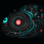
Zebrafish eye and neuromasts
This four-day-old zebrafish embryo has been modified using two mechanisms – borrowed from the fascinating worlds of bacteria and yeast – that are widely applied in genetics research. A DNA-editing technology called CRISPR/Cas9 was used to insert a gene called Gal4 next to the gene that the researchers wished to study. These Gal4 fish were then bred with special reporter fish to create fish where the gene of interest fluoresces red whenever it is activated. Here, the scientists are using these Gal4 reporter fish to study a gene expressed in the lens of the eye (the red circle in the middle of the image) the head, and cells called neuromasts (the red dots). Neuromasts form a special mechanosensory system in fish that responds to surrounding water movements and is therefore essential for a variety of behaviors, from schooling to avoiding predators. This fish’s nervous system has also been labelled for study, and is shown in blueish-green.
INGRID LEKK AND STEVE WILSON, UNIVERSITY COLLEGE LONDON
#breastcancer Twitter connections
This is a graphical visualization of data extracted from tweets containing the hashtag #breastcancer. Twitter users are represented by dots, called nodes, and lines connecting the nodes represent the relationships between the Twitter users. Nodes are sized differently according to the number and importance of other nodes they are connected with, and the thickness of each connecting line is determined by the number of times that a particular relationship is expressed within the data. The ‘double yolk’ structure at the top of the image indicates common mentions of two accounts. This area of the graph provides a graphical expression of trending data in Twitter, as it represents one tweet that was retweeted thousands of times.
ERIC CLARKE, RICHARD ARNETT AND JANE BURNS, ROYAL COLLEGE OF SURGEONS IN IRELAND
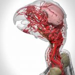
Blood vessels of the African grey parrot
This image shows a 3D reconstruction of an African grey parrot, post euthanasia. The 3D model details the highly intricate system of blood vessels in the head and neck of the bird and was made possible through the use of a new research contrast agent called BriteVu (invented by Scott Echols). This contrast agent allows researchers to study a subject’s vascular system in incredible detail, right down to the capillary level.
SCOTT BIRCH AND SCOTT ECHOLS
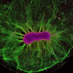
Brain-on- a-Chip
Neural stem cells have the ability to form all the different cell types found in the nervous system. Here, researchers are investigating how neural stem cells grow on a synthetic gel called PEG. After just two weeks, the stem cells (magenta) produced nerve fibers (green). These fibers grew away from the cell due to chemical gradients in the gel, teaching researchers about how their environment affects their structural organization. This work supports the Human-on- a-Chip project, which is addressing the inefficiency and cost of traditional drug testing. Researchers have devised ways of growing miniature organs on plastic chips, which they hope can be connected to represent the human body. This could be used to accurately predict the effectiveness and toxicity of drugs and vaccines and remove the need for animal testing in medical research. This image appears as a result of the partnership between Wellcome Images and the Koch Institute at MIT.
COLLIN EDINGTON AND IRIS LEE, © MASSACHUSETTS INSTITUTE OF TECHNOLOGY (MIT)
For more on this story go to; http://mashable.com/2017/03/07/2016-best-science-images-wellcome-awards/?utm_campaign=Feed%3A+Mashable+%28Mashable%29&utm_cid=Mash-Prod-RSS-Feedburner-All-Partial&utm_source=feedburner&utm_medium=feed

